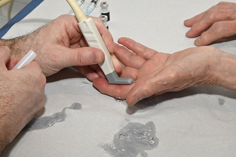
High resolution ultrasound scans of Joints
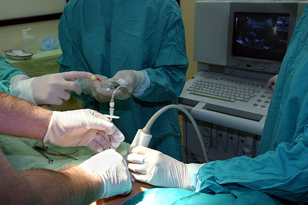
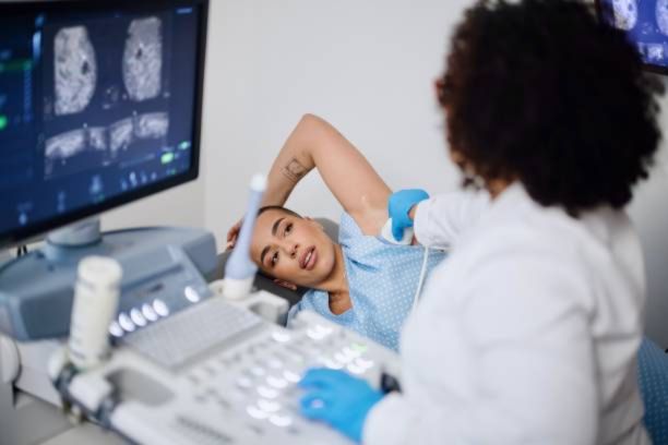
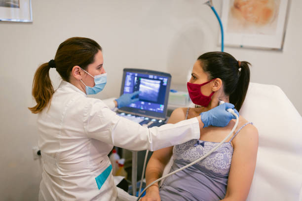
Shoulder Ultrasound
High resolution ultrasound helps in identifying various pathologies and by its dynamic evulation in real time,it can detect few specific diseases which cause pain only in few specific movements and positions, which cannot be done in other static modalities like MRI , CT , X ray etc...
Diseases that can be diagnosed:
- Frozen shoulder(adhesivecapsulitis)
- Rotater cuff muscle and tendon tears
- Supraspinatus muscle/tendon
- Infraspinatus muscle/tendon
- Teres minor muscle/tendon
- Subscapularis muscle/tendon
- Deltoid muscles muscle/tendon
- Acromioclavicular joint arthritis
- Subacromian subdeltoid bursitis
- Biceps tenosynovitis
- calcific tendonitis
- Septic arthritis
- Inflammatory arthritis
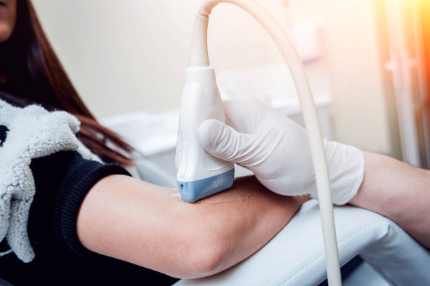
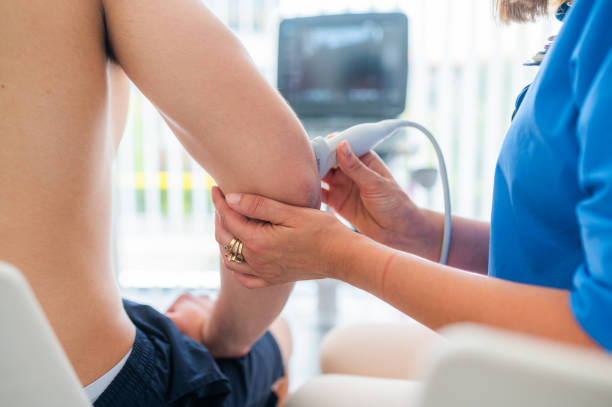
Elbow Ultrasound
Elbow Ultrasound–High-resolution ultrasound provides real-time imaging, helping identify conditions like tendinitis, bursitis, and ligament injuries that may not show up on X-rays or MRI. It can also detect fluid buildup and soft tissue inflammation, offering a dynamic view of the joint’s response during specific movements. This makes it a useful tool for diagnosing injuries from repetitive strain or sports activities.
Diseases that can be diagnosed:
- Tennis elbow(lateral epicondylitis)
- Golfers elbow(medial epicondylitis)
- Muscle and tendon tears
- biceps brachii muscle/tendon
- triceps muscle/tendon
- brachialis muscle/tendon
- pronater muscle/tendon
- supinator muscle/tendon
- Ulnar bursitis
- Nerve entrapments
- radial nerve
- ulnar nerve
- median nerve
- musculocutaneous nerve
- posterior and anterior interosseous nerve
- Ligament tears
- radial collateral ligament
- ulnar collateral ligament
- Septic arthritis
- Rheumatoid arthritis
- Gout arthritis
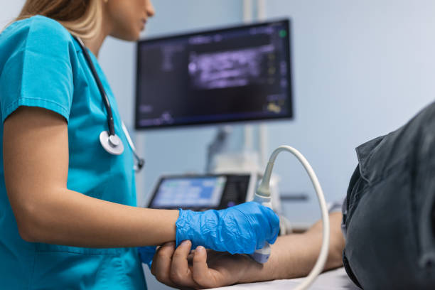
Wrist ultrasound
Wrist Ultrasound-High-resolution ultrasound aids in detecting various wrist conditions and, with its real-time dynamic evaluation, it can identify specific injuries that cause pain only during particular movements or positions, which static modalities like MRI, CT, and X-ray cannot assess.
Diseases that can be diagnosed:
- Carpal tunnel syndrome(median nerve entrapment)
- Ganglion cysts
- Muscle and tendon tears (extensor and flexor tendons)
- Dequervains tenosynovitis
- Tendons tenosynovitis (flexor and extensor group)
- TFCC ligament tears
- Proximal and distal Intersection syndromes
- Septic arthritis
- Gout arthritis
- CPPD arthritis(crystal deposition disease)
- Rheumatoid arthritis
Hand & Finger Ultrasound
Delicate and intricate,the structures of the hand and fingers require precise imaging for accurate diagnosis. Real-time ultrasound offers a dynamic view of tendons, ligaments, and joints,capturing movement-specific issues like tears and inflammation that static modalities such as MRI or CT might miss.
Diseases that can be diagnosed:
- Trigger finger(stenosing tenosynovitis)
- Tendon tears( flexor and extensor )
- Ligament tears( radial and ulnar collateral ligaments )
- Osteo arthritis
- Septic arthritis
- Gout arthritis
- Rheumatoid arthritis
- Inflammatory arthritis
- CPPD arthritis(crystal deposition disease)
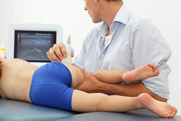
Hip Ultrasound
As the cornerstone of mobility, the hip joint demands a diagnostic approach that sees beyond the surface. Dynamic ultrasound unveils bursitis, tendinopathies, and effusions in real time, offering insights into pain or stiffness during motion—something static imaging cannot achieve.
Diseases that can be diagnosed:
- Muscle and tendon tears
- Adductor strain muscle/tendon
- hamstrings strain muscle/tendon
- Flexors muscles
- Gluteus muscles
- Iliotibial band syndrome
- Trochanteric bursitis
- Septic arthritis
- Inflammatory arthritis
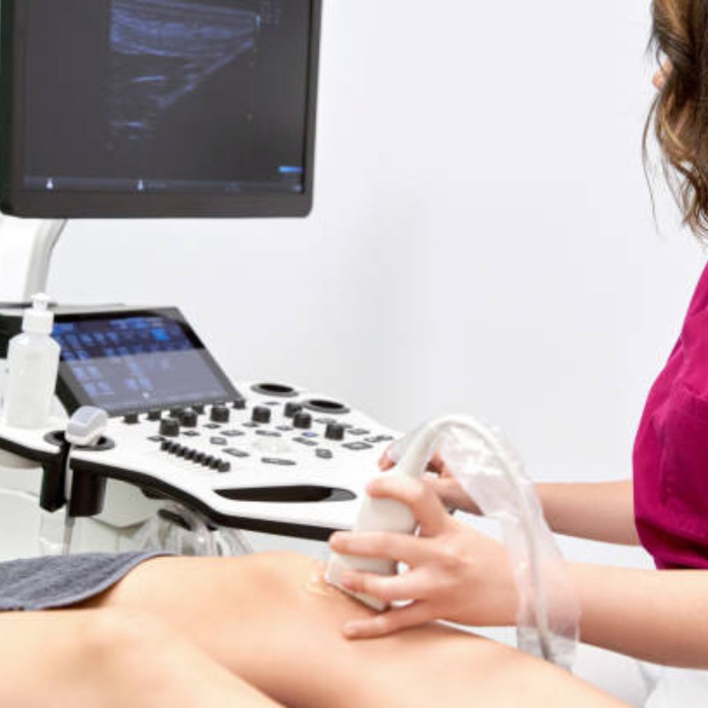
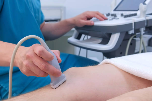
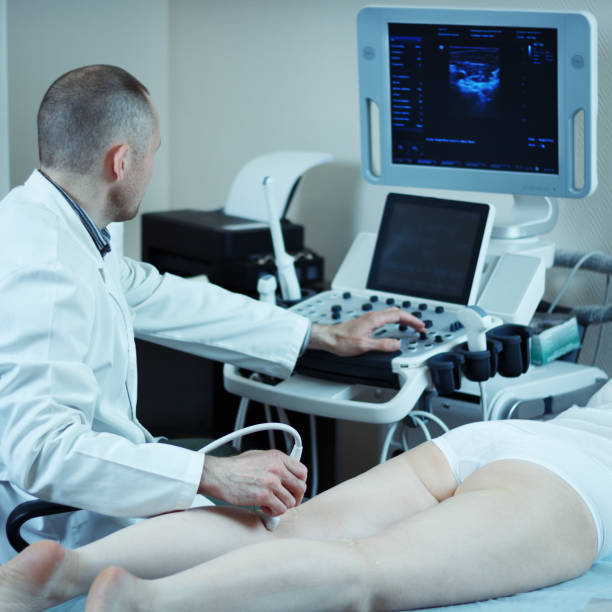
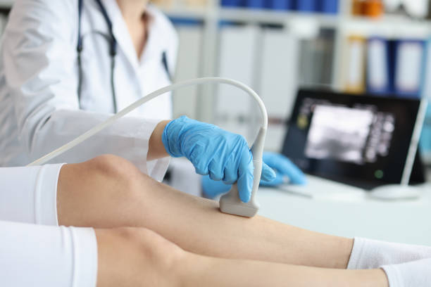
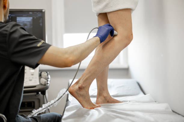
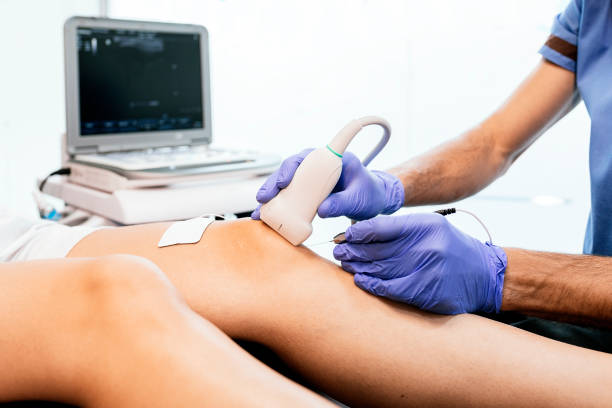
Knee Ultrasound
The knee,a pivotal joint in daily movement,benefits greatly from the dynamic evaluation of ultrasound. This advanced imaging technique reveals ligament tears, tendon injuries, and joint fluid buildup, diagnosing conditions that only manifest during specific movements.
Diseases that can be diagnosed:
- Osteoarthritis
- Muscle and tendon tears
- Quadriceps muscle/tendon
- Hamstrings muscle/tendon
- Adductors muscle/tendon
- Ligament tears
- medial and lateral collateral ligament
- anterior cruciate ligaments
- Bakers cyst
- Meniscus tears
- Fat pad impingement syndromes
- Nerve entrapments
- Sciatic nerve
- Tibial nerve
- Common peroneal nerve
- Septic arthritis
- Inflammatory arthritis
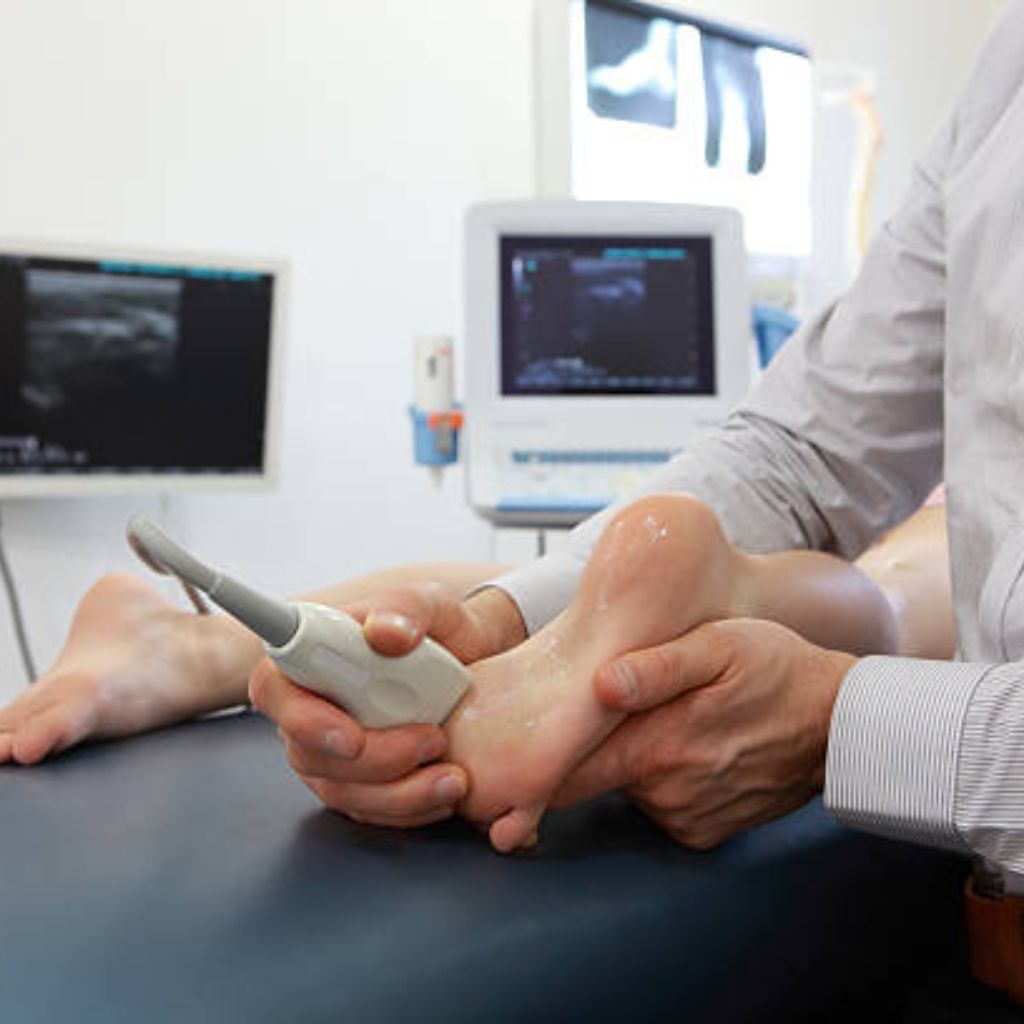
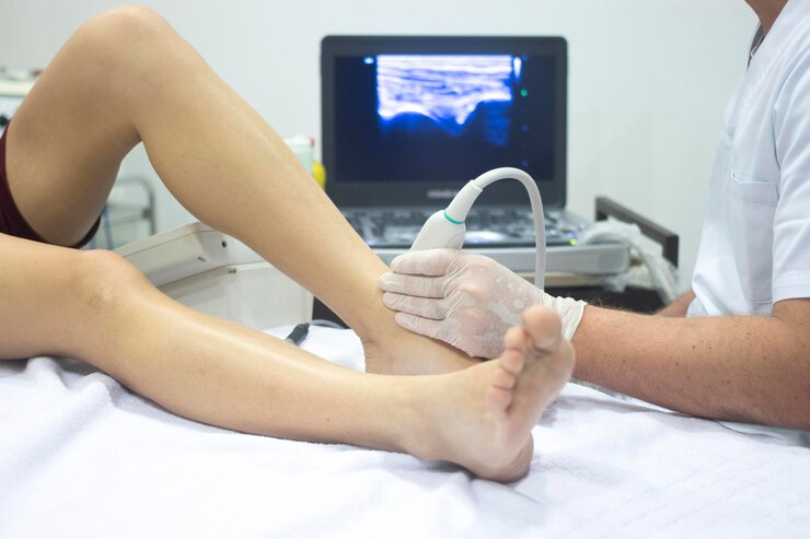
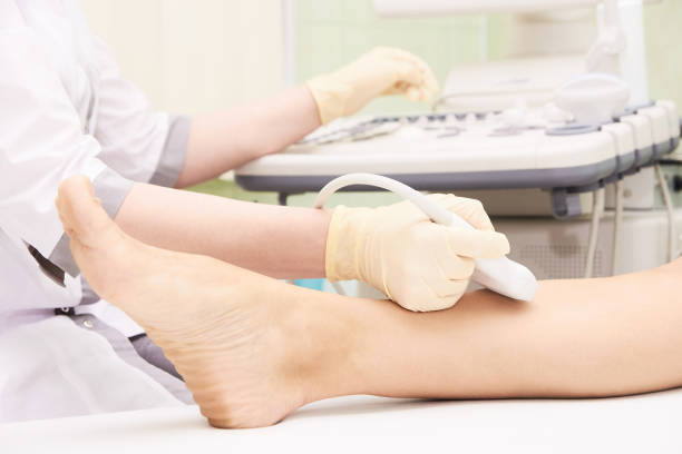
Ankle & Foot Ultrasound
High-resolution ultrasound excels in identifying soft tissue injuries, ligament sprains, and tendon tears. Its dynamic real-time evaluation uncovers movement-specific conditions that may be overlooked by MRI, CT, or X-ray.
Diseases that can be diagnosed:
- Ligament tears
- deltoid
- ATFL
- AITFL
- Spring ligament
- Muscle and tendon tears
- Tendoachillis muscle/tendon
- Tibialis posterior muscle/tendon
- Flexor and extensor muscles
- Nerve entrapments
- sural nerve
- superficial and deep peroneal nerve
- posterior tibial nerve
- lateral and medial plantar nerve
- plantar fasciitis
- retrocalcaneal and retroachillis bursitis
- Septic arthritis
- Inflammatory arthritis
- Gout arthritis
- Rheumatoid arthritis
Nerve ultrasound
Imagine uncovering the hidden causes of nerve pain and dysfunction with unparalleled precision. Nerve ultrasound offers dynamic, real-time imaging to diagnose compressions, injuries, or entrapments with accuracy. By visualizing how nerves behave during movement, it brings clarity to complex conditions, guiding targeted and effective treatments.
Diseases that can be diagnosed:
- Carpal tunnel syndrome
- Ulnar neuropathy
- Posterior interosseous nerve entrapment
- Meralgia paresthetica
- Plantar nerves entrapment
High resolution ultrasound scans of nerves
Median Nerve Ultrasound
High-resolution ultrasound provides real-time imaging to assess the median nerve for conditions like carpal tunnel syndrome. Its dynamic evaluation can detect nerve compression or entrapment during specific wrist or finger movements, offering insights beyond static imaging.
Diseases that can be diagnosed:
- Carpal Tunnel Syndrome (compression at the wrist)
- Median nerve injuries (trauma or lacerations)
- Pronator Syndrome (compression at the forearm)
Ulnar Nerve Ultrasound
Real-time ultrasound enables detailed assessment of the ulnar nerve, identifying entrapment or compression at sites like the elbow or wrist. Its dynamic capabilities help uncover movement-induced abnormalities missed by MRI or CT.
Diseases that can be diagnosed:
- Nerve entrapments or swelling (e.g., tumors, cysts)
- Guyon’s Canal Syndrome (compression at the wrist)
- Cubital Tunnel Syndrome (compression at the elbow)
Radial Nerve Ultrasound
High-resolution ultrasound effectively visualizes the radial nerve, aiding in the diagnosis of injuries or compression syndromes. Its real-time imaging allows dynamic evaluation during arm or wrist movement, providing insights not possible with static modalities.
Diseases that can be diagnosed:
- Radial Tunnel Syndrome (compression near the elbow)
- Wartenberg’s Syndrome (superficial radial nerve entrapment)
- Radial nerve injuries (fractures or trauma)

Screening of All Upper Limb Nerves
Comprehensive high-resolution ultrasound screening evaluates all major upper limb nerves, including the median, ulnar, and radial nerves. Real-time dynamic assessment identifies movement-specific abnormalities and provides a complete diagnostic view for nerve-related conditions.
Diseases that can be diagnosed:
- Generalized neuropathies (diabetes, systemic diseases)
- Nerve injuries due to trauma, fractures, or surgical complications
- Nerve entrapments (median, ulnar, radial) at multiple levels
Common Peroneal Nerve Ultrasound
As a key nerve for lower limb mobility, the common peroneal nerve is prone to compression or injury, especially around the knee. Dynamic ultrasound provides detailed visualization, capturing abnormalities like entrapment or swelling during motion, which static imaging techniques may miss.
Diseases that can be diagnosed:
- Nerve injuries due to trauma or fractures (e.g., proximal tibia or fibula)
- Nerve entrapment caused by masses (tumors, cysts, lipomas)
- Neuropathies associated with diabetes or systemic conditions
Sciatic Nerve Ultrasound
The sciatic nerve, the largest in the body, can be affected by trauma,entrapment,or inflammation. Ultrasound offers a comprehensive, real-time evaluation, allowing clinicians to identify conditions impacting the nerve along its course with precision and clarity.
Diseases that can be diagnosed:
- Sciatic Neuropathy (compression or injury in the gluteal or thigh region)
- Traumatic injuries from fractures or hip dislocations
- Inflammatory neuropathies or nerve swelling
Plantar Nerves Ultrasound
Essential for diagnosing foot pain and dysfunction, plantar nerve ultrasound reveals conditions like entrapments or nerve compressions that affect mobility. Its real-time imaging capability highlights abnormalities during specific movements, aiding in targeted diagnosis and treatment.
Diseases that can be diagnosed:
- Neuromas (e.g., Morton’s neuroma)
- Tarsal Tunnel Syndrome (compression of the tibial nerve or its branches)
- Plantar fasciitis with associated nerve involvement
Screening of Lower Limb Nerves
For a thorough assessment of lower limb nerve health, ultrasound screening offers dynamic, real-time imaging of major nerves, including the sciatic, common peroneal, and plantar nerves. This comprehensive approach ensures accurate diagnosis of movement-specific abnormalities and nerve-related conditions
Diseases that can be diagnosed:
- Generalized neuropathies (e.g.,due to diabetes or systemic diseases)
- Post-traumatic or post-surgical nerve injuries
- Compression neuropathies at multiple levels (e.g., lumbar plexus)
Brachial plexus ultrasound
High-resolution imaging technique used to visualize the brachial plexus, a network of nerves that originates from the cervical spine and controls motor and sensory functions of the shoulder, arm, and hand. It is a non-invasive tool that helps assess the structure, continuity, and pathological changes in the brachial plexus and surrounding tissues.
Diseases that can be diagnosed:
- Congenital Abnormalities
- Post-Surgical or Iatrogenic Injuries
- Tumors and Masses
- Inflammatory Conditions
- Neuropathies
- Nerve Compression Syndromes
- Traumatic Injuries
General Ultrasound scans
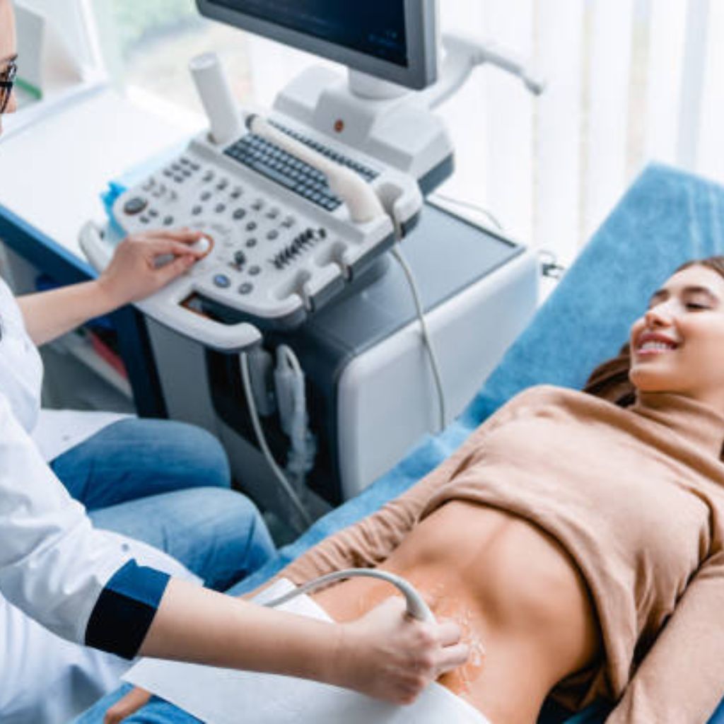
Ultrasound Abdomen and Pelvis
Ultrasound of the abdomen and pelvis provides a non-invasive, detailed view of internal organs such as the liver, kidneys, bladder, uterus, and ovaries. This safe and painless procedure aids in diagnosing conditions like organ enlargement, fluid accumulation, or abnormalities in the urinary or reproductive systems.
Diseases that can be diagnosed:
- Liver Diseases
- Fatty liver
- cirrhosis
- hepatomegaly
- liver tumors
- abscesses
- Gallbladder Diseases
- Gallstones
- cholecystitis
- polyps
- sludge
- Kidney and Urinary Tract Diseases
- Kidney stones
- hydronephrosis
- cysts
- bladder abnormalities
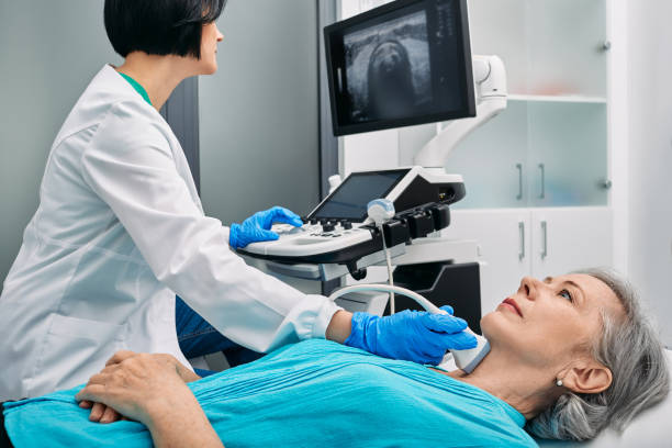
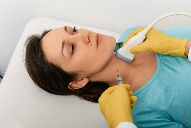
High-Resolution Ultrasound of Neck
A high-resolution neck ultrasound offers detailed imaging of the thyroid gland, lymph nodes, salivary glands, and other soft tissues. This advanced diagnostic tool is essential for evaluating thyroid nodules, gland enlargement, or lymphatic abnormalities, providing accurate results in real-time.
Diseases that can be diagnosed:
- Thyroid Diseases
- Thyroid nodules
- goiters
- thyroiditis
- Lymph Node Abnormalities
- Enlarged lymph nodes
- lymphadenitis
- Parathyroid Disorders
- Parathyroid adenomas or hyperplasia
High-Resolution Ultrasound of Soft Parts / Local Region
This ultrasound specializes in evaluating localized areas like muscles, tendons, ligaments, and superficial soft tissues. It is particularly helpful for identifying cysts, abscesses, or soft tissue injuries with unmatched precision and real-time imaging capabilities.
Diseases that can be diagnosed:
- Soft Tissue Infections
- Cellulitis
- myositis
- Tendon and Muscle Injuries
- Tears
- sprains
- Superficial Masses
- Lipomas
- sebaceous cysts
Color Doppler Studies
Upper Limb / Lower Limb Arterial Doppler
This study evaluates blood flow in the arteries of the upper and lower limbs. It helps identify conditions such as arterial blockages, stenosis, or peripheral vascular disease, providing vital insights for timely treatment.
Diseases that can be diagnosed:
- Peripheral Arterial Disease (PAD)
- Stenosis
- occlusions
- Aneurysms
- Arterial dilations or pseudoaneurysms
- Arterial Insufficiency
- Ischemia
- reduced blood flow
Upper Limb / Lower Limb Venous Doppler
Venous Doppler imaging assesses blood flow in the veins, detecting issues like deep vein thrombosis (DVT), varicose veins, or venous insufficiency. It is an invaluable tool for identifying circulatory disorders and planning appropriate interventions.
Diseases that can be diagnosed:
- Chronic Venous Insufficiency
- Varicose veins
- venous reflux
- Venous Obstructions
- Compression syndromes
- thrombus
Carotid Doppler / Neck Vessel Doppler
Carotid Doppler imaging evaluates blood flow in the carotid arteries and other neck vessels. This test is crucial for detecting plaques, stenosis, or blood flow irregularities that may lead to stroke or other vascular complications.
Diseases that can be diagnosed:
- Carotid Artery Stenosis
- Atherosclerotic plaques
- narrowing
- Vascular Insufficiency
- Reduced blood flow to the brain
- Arterial wall injuries
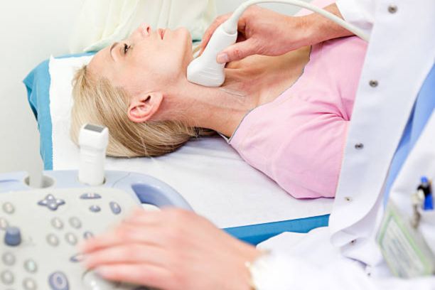
Image guided Interventional procedures
Ultrasound-Guided Fine Needle Aspiration Cytology (FNAC)
Ultrasound-guided FNAC is a minimally invasive procedure used to obtain tissue or cell samples for diagnostic purposes. It is often employed to evaluate lumps or masses, helping determine the underlying cause and guiding further management.
Diseases that can be diagnosed:
- Thyroid Nodules
- Benign or malignant diagnosis
- Lymph Node Biopsies
- Identification of metastases
- infections
- Salivary Gland Lesions
- Tumors or inflammatory conditions
Ultrasound-Guided Biopsy
This precise procedure uses ultrasound to guide the extraction of tissue samples from a specific site, such as the liver, lymph nodes, or breast. It plays a critical role in diagnosing cancers, infections, or inflammatory conditions.
Diseases that can be diagnosed:
- Liver and Kidney Lesions
- Tumor diagnosis or grading
- Breast Masses
- Malignant versus benign differentiation
- Deep-Seated Masses
- Retroperitoneal
- pelvic
- thoracic masses
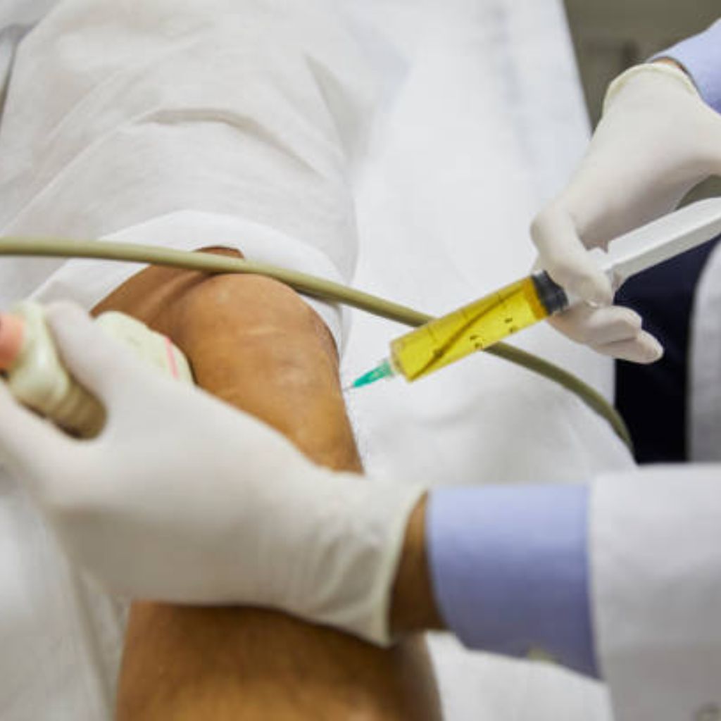
Ultrasound-Guided Fluid Aspiration
This procedure is performed to remove fluid from areas such as joints, cysts, or abscesses. Ultrasound guidance ensures safe and accurate fluid removal, relieving symptoms and aiding in the diagnosis of infections or other conditions.
Diseases that can be diagnosed:
- Abscess Drainage
- Liver
- kidney
- soft tissue abscesses
- Cyst Aspiration
- Ovarian
- hepatic
- pancreatic cysts
- Joint Effusions
- Septic arthritis
- synovial fluid collection
Ultrasound-Guided Pigtail Catheter Insertion
A pigtail catheter is inserted under ultrasound guidance to drain fluid or abscesses from the chest, abdomen, or other areas. This minimally invasive technique is essential for managing pleural effusion, ascites, or localized infections effectively..
Diseases that can be diagnosed:
- Pleural Effusion Drainage
- Malignant or infectious effusions
- Ascitic Fluid Drainage
- Cirrhosis-related or malignant ascites
- Pericardial Effusion Drainage
- Relieving cardiac tamponade